Meniscus Tears
Specialized care for meniscus injuries in athletes and active individuals. Treatment is focused on preserving knee function, protecting long-term joint health, and helping patients safely return to sports and the activities they love.
Understanding Meniscus Tears
Anatomy of the Knee
The knee is one of the largest and most complex joints in the body. It is formed by three bones: the femur (thigh bone), tibia (shin bone), and patella (kneecap). Together, these structures allow the knee to bend, straighten, absorb force, and support the body during daily activities and sports.
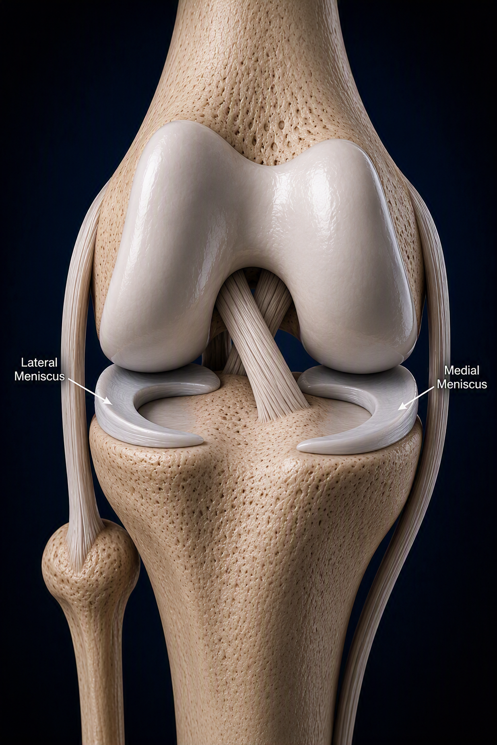
Between the femur and tibia are two important shock absorbers called the menisci. Each knee contains a medial (inner) meniscus and a lateral (outer) meniscus. These structures help distribute load, protect the joint cartilage, provide stability, and absorb shock during movement.
Because of their important role in knee function, preserving the meniscus whenever possible is a major goal of treatment.
Understanding Meniscus Tears
What is a Meniscus Tear?
A meniscus tear occurs when the cartilage within the knee becomes injured. Meniscus tears may occur as a result of a sports injury, twisting injury, or normal age-related degeneration.
Meniscus tears commonly occur:
During sports and athletic activity
Following twisting injuries
In combination with ACL tears
As part of the aging process
Anatomy of the Knee
The knee is one of the largest and most complex joints in the body. It is formed by three bones: the femur (thigh bone), tibia (shin bone), and patella (kneecap). Together, these structures allow the knee to bend, straighten, absorb force, and support the body during daily activities and sports.
Between the femur and tibia are two important shock absorbers called the menisci. Each knee contains a medial (inner) meniscus and a lateral (outer) meniscus. These structures help distribute load, protect the joint cartilage, provide stability, and absorb shock during movement.
Because of their important role in knee function, preserving the meniscus whenever possible is a major goal of treatment.
Common Symptoms of a Meniscus Tears
The knee is one of the largest and most complex joints in the body. It is formed by three bones: the femur (thigh bone), tibia (shin bone), and patella (kneecap). Together, these structures allow the knee to bend, straighten, absorb force, and support the body during daily activities and sports.
Between the femur and tibia are two important shock absorbers called the menisci. Each knee contains a medial (inner) meniscus and a lateral (outer) meniscus. These structures help distribute load, protect the joint cartilage, provide stability, and absorb shock during movement.
Because of their important role in knee function, preserving the meniscus whenever possible is a major goal of treatment.
Types of Meniscus Tears
Anterior/Posterior Horn Tears:
These are the most common and often occur in younger patients or athletes. The posterior horn is the back part of the meniscus and is crucial for knee stability. Tears in this region are often repaired when possible, especially in combination with ACL reconstruction.
Body Tears:
Tears in the central portion of the meniscus may be repairable depending on their size, location, and tissue quality. If the tear is in an area with poor blood supply, it may require partial meniscectomy (trimming of the torn portion).
Radial Tears:
These are perpendicular tears that cut across the meniscus and disrupt its ability to distribute load. Depending on the location, radial tears may be repairable, particularly if they are near the root or in a vascular zone.
Bucket-Handle Tears:
A type of tear where a portion of the meniscus flips into the joint, often causing locking or catching. These typically require urgent surgical repair, especially if the knee is locked.
Meniscus Root Tears:
These involve a tear at the root (attachment site) of the meniscus to bone and can mimic the effects of a total meniscectomy if not treated. Root tears are often repaired surgically to restore normal knee mechanics and prevent early cartilage damage.
Treatment Options
Non-Surgical Treatment
Many meniscus tears can be successfully treated without surgery.
Treatment may include:
Physical therapy
Anti-inflammatory medications
Activity modification
Corticosteroid injections
Strengthening programs
Gradual return to activity
Depending on the tear type, patients can experience significant symptom improvement and can avoid surgery altogether.
When Is Surgery Recommended?
Surgery may be recommended for patients with:
Persistent pain despite rehabilitation
Mechanical symptoms such as locking or catching
Displaced meniscus tears
Meniscus root tears
Associated ACL injuries
Limitations in sports or daily activities
Whenever possible, preserving the meniscus is preferred because it plays an important role in protecting the long-term health of the knee.
Meniscus Surgery
Depending on the location and pattern of the tear, surgery may involve:
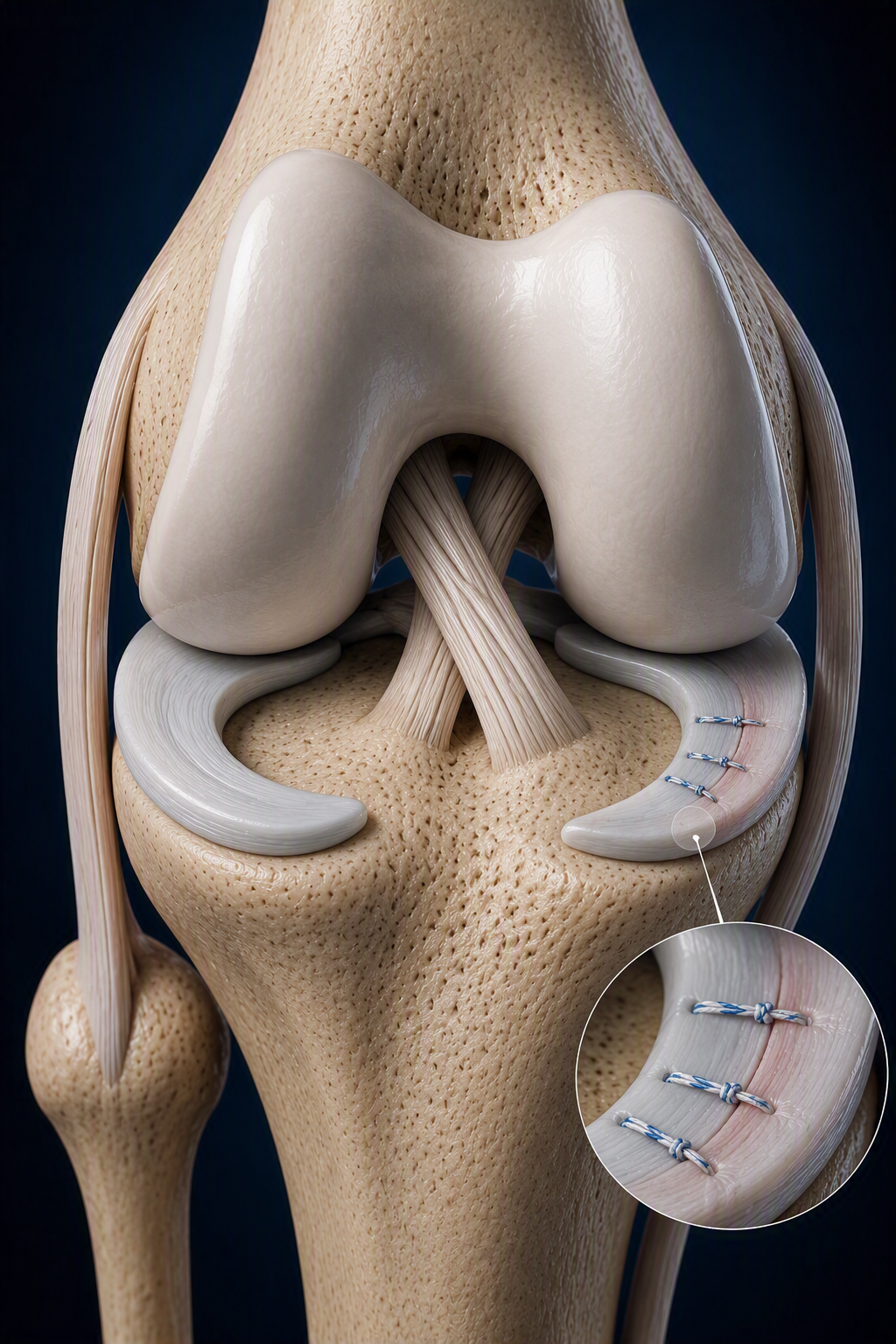
The torn meniscus is repaired using sutures to allow healing. Meniscus repair is preferred whenever possible because it preserves the native cartilage and helps protect the knee long-term.
Meniscus Repair
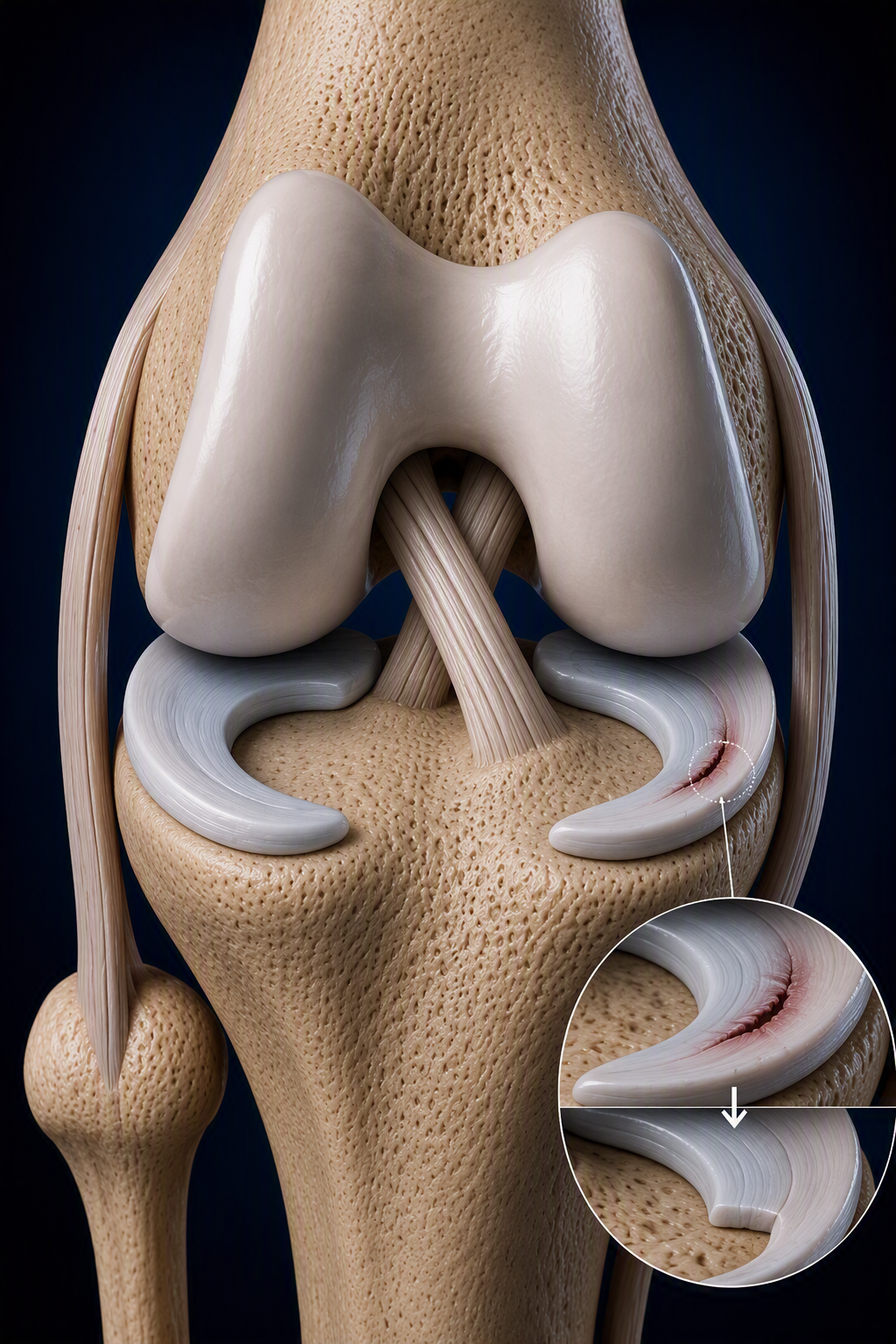
When a tear is not repairable, the unstable portion of the meniscus may be trimmed while preserving as much healthy meniscus tissue as possible.
Partial Meniscectomy
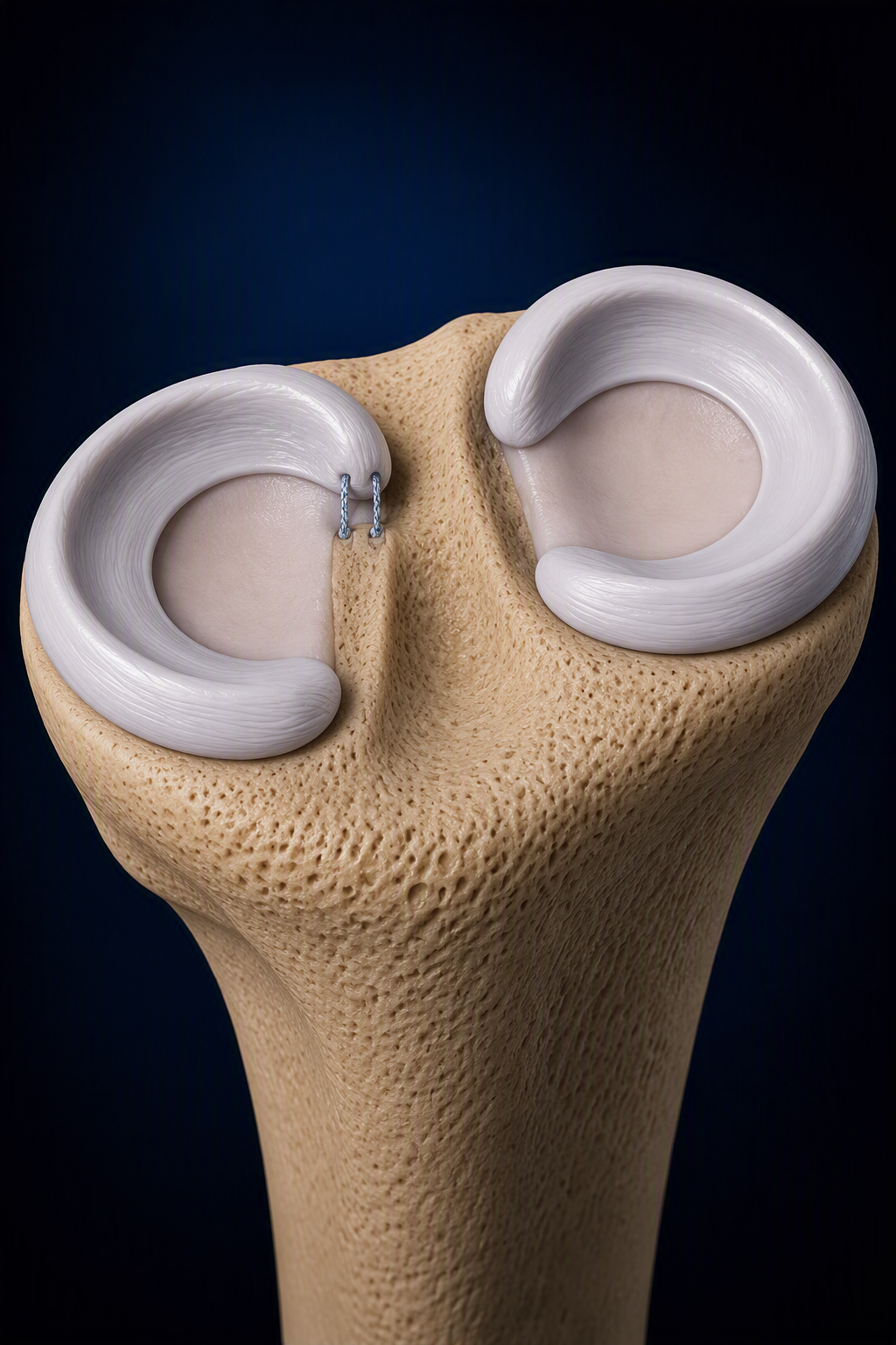
Specialized repair techniques are used to restore meniscus root function and re-establish normal knee biomechanics.
Meniscus Root Repair
General Meniscus Tear
Frequently Asked Questions
-
The meniscus acts as a shock absorber within the knee and helps distribute load, provide stability, and protect the joint cartilage.
-
Some small tears may become asymptomatic over time. However, healing depends on the location of the tear and its blood supply.
-
No. Many meniscus tears can be successfully treated with physical therapy, activity modification, and other non-operative measures.
-
Whenever possible, meniscus preservation is preferred because it helps protect the cartilage and long-term health of the knee.
-
Repairability depends on the tear pattern, location, tissue quality, blood supply, age, and activity level.
-
Most patients are able to return to sports and physical activity following appropriate treatment and rehabilitation.
-
Common symptoms include pain, swelling, catching, locking, stiffness, and difficulty with twisting or squatting activities.
-
A bucket-handle tear is a displaced meniscus tear in which a portion of the meniscus flips into the joint and may cause the knee to lock.
-
A root tear occurs where the meniscus attaches to bone. These injuries significantly affect knee mechanics and often require surgical treatment.
-
Some tears may remain stable, while others can continue to cause pain, mechanical symptoms, or additional cartilage damage over time.
-
Yes. Meniscus tears frequently occur alongside ACL injuries, particularly in athletes and active individuals.
-
The meniscus helps protect the cartilage surfaces of the knee. Preserving as much healthy meniscus tissue as possible may reduce the risk of future arthritis and improve long-term knee function.
Patient Resources
Meniscus Surgery
Frequently Asked Questions
-
If you had a meniscectomy, you will not need a brace
If you had a meniscal repair, you can take the brace off for showering, dressing, and to do your exercises with physical therapy. You can also remove the brace while icing. However, you must wear the brace at all other times, including sleeping and when walking. The brace will be worn for at least 6-12 weeks.
-
The ability for someone to resume driving after surgery is seldom a medical question, but more often a legal question. Driving with any form of a brace on may be interpreted as driving while impaired. It is the responsibility of all licensed drivers to drive safely at all times no matter what their permanent or temporary impairment may be.
-
Avoid air travel for a minimum of two weeks after surgery on the lower extremity to decrease your risk of blood clots. If air travel is necessary, please inform the office as we will increase your dose of aspirin to 325mg twice a day for 5 days. Also, if air travel is necessary either walk frequently or do ankle pump exercises (flex your foot up and down repeatedly) in your seat every hour. This squeezes the blood vessels, helping to prevent the formation of blood clots. Stay well hydrated as dehydration causes the blood to thicken. Avoid alcohol consumption as alcohol can cause dehydration.
-
We will need to see you for a follow-up appointment around 10-14 days after your surgery. Dr. Danaher will then assess your recovery to determine the timing of additional appointments. Depending on your procedure, you will be seen at 2 weeks, 6 weeks, 3 months, 6 months, 9 months and one year after surgery. The timing of further visits will be determined by Dr. Danaher.
-
This is variable, of course, but we provide you with an appropriate amount of narcotic pills to keep you comfortable until your 1st post-operative visit. You should take these pills as needed. You can take 1-2 pills every 4-6 hours. You can also take 600mg of Advil, Aleve, Motrin, or ibuprofen in conjunction with your pain medication.
-
Most patients receive a regional block unless there is a contraindication to it. Further anesthesia is determined by your anesthesiologist. Your anesthesiologist will speak with you before the procedure and address any of your questions or concerns
-
This really depends on the individual patient specifically with respect to job demands (labor vs desk job). Desk jobs may return to work within days and labor jobs typically around 3 months
-
If you had a meniscectomy, patients typically return to sports in about 6-8 weeks.
If you had a meniscal repair, patients typically return to sports in about 6 months
However, you will be able to participate in certain activities in a progressive manner throughout your recovery. These activities depend on your progress and timeframe of recovery and will be determined by your therapist and/or doctor.
-
If you had a meniscectomy, Physical Therapy is typically done for 4-6 weeks
If you had a meniscus repair, The typical therapy program will be a minimum of 6 months. The 1st phase of therapy will be designed to safely regain your range of motion while the 2nd phase of therapy will be directed at regaining function, strength, and endurance.
-
When you do not have any pain, inflammation, or swelling, you may discontinue cryotherapy. If you experience any discomfort or inflammation after exercising or returning to activity, resume your cryotherapy immediately. Even when you do not have immediate symptoms, you may utilize cryotherapy preventively to avoid any residual symptoms. Use for 3-7 days, 3-5 times per day for 30 minutes to 1 hour each session.
-
You should plan to wear loose, comfortable clothing and supportive, stable shoes. Slip on shoes are easier to get into after surgery.
-
Yes. But other valuables (Laptops, iPads, iPods, Jewelry, etc) should be left at home.
-
Do not assume it will heal on its own. Please call Dr. Danaher's office right away to determine if you need an evaluation.
-
Any signs of infection should be reported immediately – these include increased drainage (usually thick, cloudy, not liquid secondary to the arthroscopy), redness, increased warmth, and fevers (T > 101.8).